Hyperspectral imaging for visualizing molecular tissue signatures

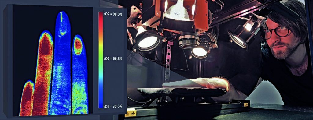
Traditional optical imaging techniques are based on monochromatic or RGB imaging and do not capture the entire spectrum, which limits many diagnostic procedures to simple imaging. Hyperspectral imaging (HSI), on the other hand, opens up a wide range of new applications. For example, HSI enables precise monitoring of perfusion, i.e., the blood flow to organs and wounds, during surgical procedures. Currently, parameters from Doppler sonography and pulse oximetry are used for monitoring, but these cannot be reliably applied to open wounds and organs. As a result, the assessment of tissue damage is often based on the subjective judgment of the attending physician. In order to make an accurate diagnosis, tissue must be removed from patients and then analyzed, which makes real-time monitoring impossible.
Hyperspectral imaging could bring about a paradigm shift here. It offers a patient-friendly solution, as it can be easily integrated into routine diagnostics. Clinically relevant information is captured at a high data rate and with chemical specificity and analyzed using AI methods. The data obtained can also be visualized using augmented reality in the form of data cubes to display it together with the tissue being examined.
The aim of the project is to develop a modular HSI system that can be used both in the perioperative phase and endoscopically. The integration of suitable AI models should enable the visualization of molecular tissue signatures using hyperspectral imaging. The project aims to transfer the technology into clinical research, with initial tests planned in cooperation with Jena University Hospital.
The main project to which HyperVismo belongs can be found here.

Andreas Kleiber