Nov 2020 – Jan 2022
Project assignment
Calvin Kreft & Konstantin Gramatte
Without light, vision as we know it would be inconceivable. But what other applications are possible that are not immediately obvious to us? Optical coherence tomography (OCT) is a high-resolution method for the multidimensional visualization of structures below what we can see.
The student project involved the development of such an OCT set-up, which was intended to explain the complex implementation of the imaging process to the students in a clear and practical way and enable a (playful) “hand-on experience” with this system. The modular system for optical systems from the open source project “Bionanoimaging openUC2” on GitHub served as a guide. The modular simplification of the structure has a major advantage here: it leads to the avoidance of (operating) errors and thus to a reduction in maintenance costs, as replacements could be provided in the shortest possible time in the event of failures.
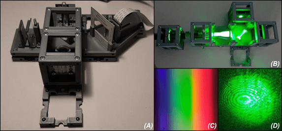
The basic idea is simple; different components of the optical system were to be easily attached to a base plate with recessed magnetic spheres within modular cubes. At the heart of the setup was the Michelson interferometer, which is used to create interference phenomena, i.e. the interaction of light with itself (see photo). Various optical elements, such as a beam splitter or lenses made of acrylic glass, a broadband light source and a spectrometer then formed the complete OCT system. Spectrometers are opto-mechanical components for splitting light and displaying a spectrum. The detector, a Raspberry Pi, captured the spectrally split light from a smartphone LED and then converted the information into intensity profiles (A-scan) of the measured depth composition.
The project was subsequently continued as part of a student research project.
Jun 2021 – Dec 2021
Funded students research project
Calvin Kreft & Konstantin Gramatte

Optical coherence tomography (OCT) is an imaging method for generating two- and three-dimensional images of depth profiles in organic and non-organic media, similar to ultrasound imaging. Due to the high axial resolution of around 0.5 μm to 15 μm and a relatively high penetration depth of 1-3 mm, depending on the wavelength used, this method offers a wide range of applications, including in medicine and materials analysis. Ophthalmology in particular benefits from the technological advances of OCT thanks to the high-resolution imaging, for example when examining the anterior and posterior segment of the eye.
Building on the previous Master’s project, the project comprised the development and construction of an OCT system, which was intended to explain the implementation of the imaging procedure to the students in a clear and practical way and enable a “hand-on experience” with this procedure. Based on the open-source bionanoimaging openUC2 project and the progress made so far, a basic setup was already available. In order to improve the signal quality, the optical setup was extended and numerous elements were optimized. Furthermore, the students developed an adapted algorithm for converting the spectral information into two-dimensional gray-scale images (B-scan).
In the course of the research, however, numerous unavoidable complications in the low-cost optical setup became apparent, some of which required cost-intensive and complex solutions that exceeded the time frame. These included the necessary use of a broadband laser, high-precision translation axes and a detector with a much higher signal-to-noise ratio.

